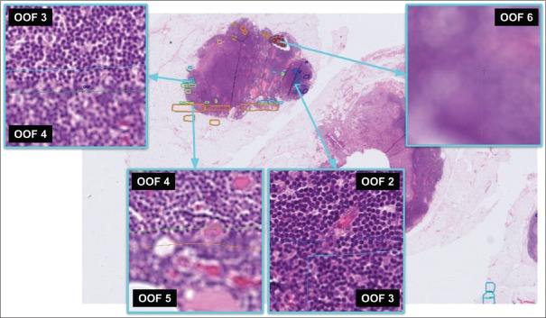
Figure 6.
Examples of OOF annotations by pathologist. The pathologist identified, delineated and graded the regions highlighted in this lymph node from a colon cancer case (scanned at × 40 on Leica AT2). Colors from the jet palette indicate the manual OOF annotation grade, ranging from 0 (in-focus, dark blue) to 6 (strong OOF, red). OOF: Out-of-focus

