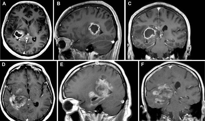
FIGURE 5.
Postcontrast axial A, sagittal B, and coronal C MRI of the brain demonstrating a glioblastoma originating in the right lateral thalamus and expanding beyond the thalamus into the periatrial region with compressing of the posterior limb of the internal capsule. Progression D-F into the fornix, parahippocampal gyrus, and mesial structures is appreciated 6 mo following initial diagnosis and adjuvant chemoradiation therapy.

