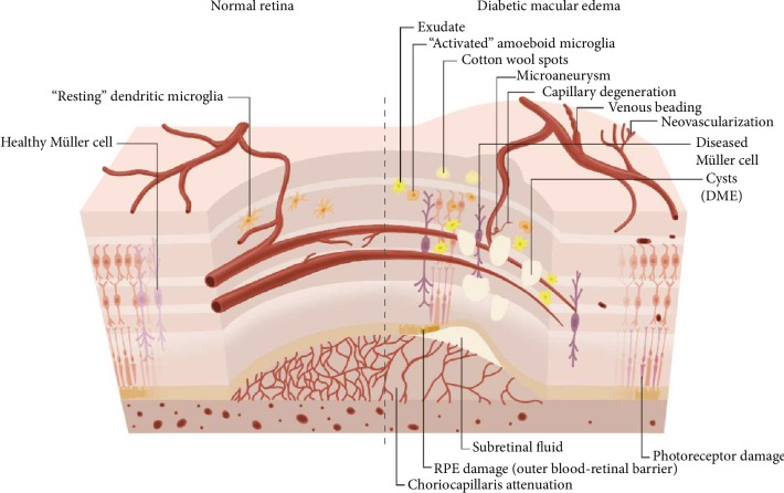
Figure 1.
Schematic illustration of the BRB of a healthy retina compared with DME. DME exhibits vascular changes including microaneurysms, capillary degeneration, venous beading, neovascularization, associated with activated microglia, Müller cell swelling, retinal pigment epithelium RPE damage, and choriocapillaris attenuation. Breakdown of BRB results in subretinal fluid and retinal cysts.

