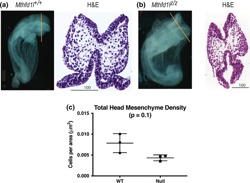
FIGURE 4.
Head mesenchyme density in paraffin sections in 7–13 somite stage embryos. Representative transverse paraffin-embedded sections of Mthfd1l+/+ (wild-type) (a) and Mthfd1lz/z (nullizygous) (b) embryos were stained with hematoxylin and eosin (H&E). Straight line in the whole embryos indicates the level of sections. Scale bars indicate microns. (c) Total head mesenchyme density. n = three embryos in each genotype with values from three sections averaged per embryo. p value was calculated by Mann–Whitney U test

