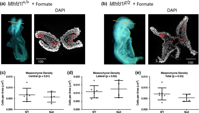
FIGURE 5.
Maternal formate supplementation improves head mesenchyme density in Mthfd1l mutant embryos at 7–13 somite stages. (a, b) Representative transverse cryosections of the cranial region of Mthfd1l+/+ (wild-type) and Mthfd1lz/z (nullizygous) embryos were stained with DAPI for nuclei. Straight line in the whole embryos indicates the level of sections. Scale bars indicate microns. (c–e) Head mesenchyme density is quantified for central and lateral areas (boxed regions in DAPI stained sections), as well as the total mesenchyme. n = six embryos in formate-supplemented wild type and four embryos in formate-supplemented nullizygotes with values from two to four sections averaged per embryo.p values were calculated by Mann–Whitney U test

