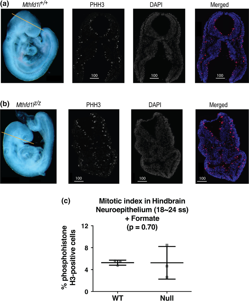
FIGURE 7.
Maternal formate supplementation improves reduced proliferation in neuroepithelium of Mthfd1l mutant embryos at 18–24 somite stages, at the time of completion of neural tube closure. Straight line in whole embryos (a, b) indicates the level of sections. Sections were stained for phosphohistone H3 (PHH3) and DAPI for nuclei. In the merged images, PHH3 staining is red and DAPI staining is blue. (a) Representative sections from Mthfd1l+/+ (wild-type) embryo. (b) Representative sections from Mthfd1lz/z (nullizygous) embryo. Scale bars indicate microns. (c) The mitotic index (% phosphohistone H3-positive cells) in neuroepithelium was quantified at the developing hindbrain. n = three embryos for both genotypes and the plot represents mean value of three to four sections per embryo. Error bars represent standard deviation, and p value was calculated by Mann–Whitney U test

