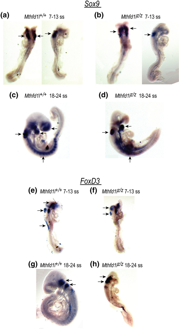
FIGURE 9.
Deletion of Mthfd1l does not affect neural crest cell specification during neural tube closure. Sox9 and FoxD3 expression were visualized by whole mount in situ hybridization. (a–d) Sox9 expression in premigratory and migrating neural crest cells in the developing forebrain, hindbrain, otocyst, head mesenchyme, somites, and the branchial arches. (e–h) FoxD3 expression in premigratory neural crest cells in the dorsal neural tube, otocyst, surface ectodermal cells overlying the spinal cord, trigeminal ganglion and vestibular-acoustic ganglion (arrows)

