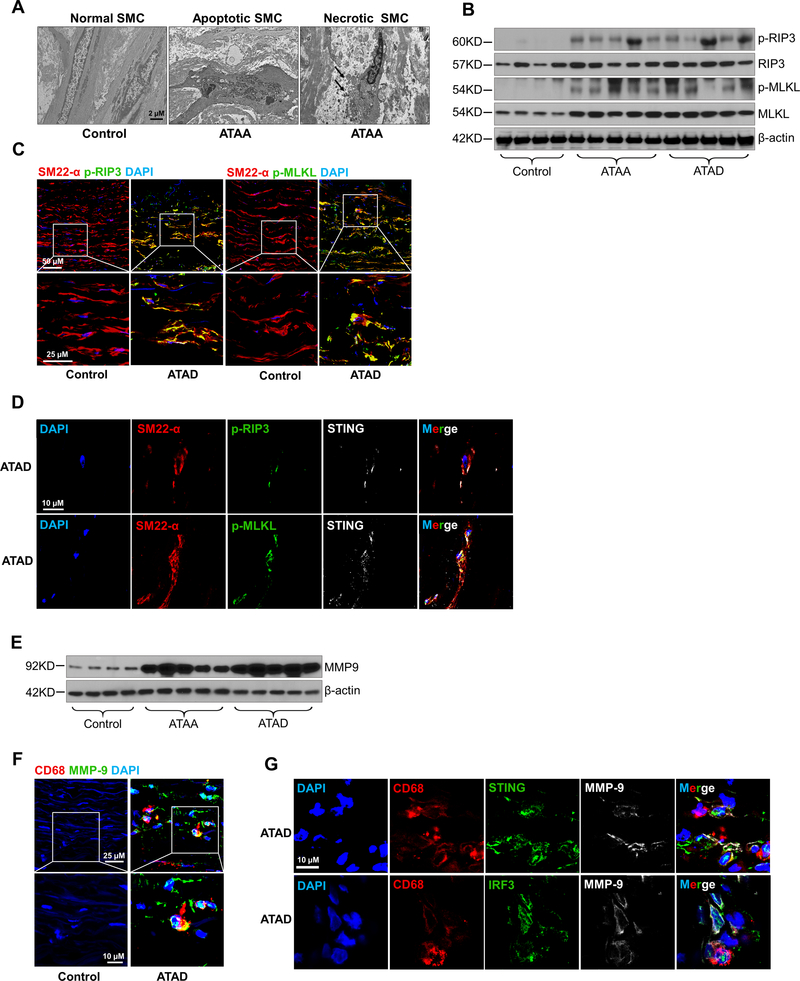
Figure 7. Association of STING activation with smooth muscle cell (SMC) injury and MMP-9 production in human sporadic ascending thoracic aortic aneurysm and dissection (ATAAD) tissues.
A, Electron microscopy images of ascending thoracic aortic aneurysm (ATAA) patient tissue showing apoptotic SMC death with nuclear fragmentation, chromatin condensation, and blebbing of the cell membrane and necrotic SMC death with swollen nuclei, mitochondria, and endoplasmic reticulum. B, Western blot analysis showing increased phosphorylation of RIP3 (p-RIP3) and MLKL (p-MLKL) in the aortic wall of ATAAD patients. C, Immunostaining showing increased p-RIP3 and p-MLKL levels in SMCs (SM22-α) of ascending thoracic aortic dissection (ATAD) tissues. Insets show a higher-magnification view. D, Immunostaining showing the colocalization of STING with p-RIP3 and p-MLKL in the SMCs (SM22-α) of ATAD tissues. E, Western blot analysis showing increased MMP-9 levels in ATAAD patient tissues. F, Immunostaining showing increased MMP-9 expression in macrophages (CD68) of diseased tissues from patients with ATAD and G, the colocalization of MMP-9 with STING and IRF3 in macrophages (CD68) of diseased tissues from patients with ATAD. Insets show a higher-magnification view.

