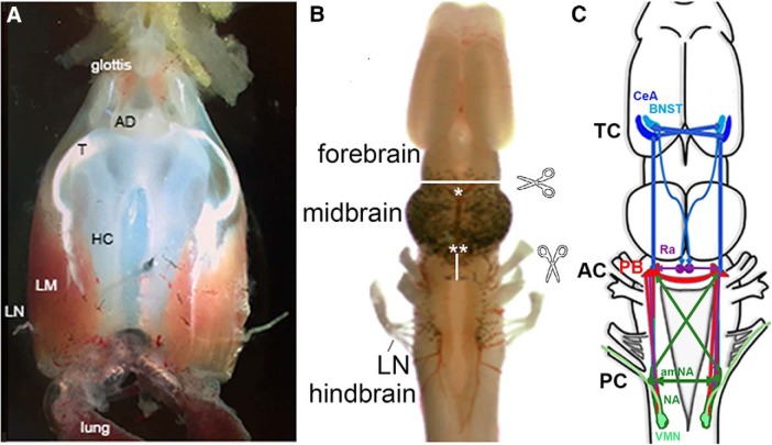
Figure 2.
A, The ex vivo larynx, dorsal view; anterior is up. The larynx communicates with the oral cavity via the glottis anteriorly and with the lungs posteriorly. The sound producing elements are the arytenoid disks (ADs), which connect via a tendon (T) to intrinsic laryngeal muscles (LM), which wrap around the hyaline cartilage (HC). When the laryngeal nerves (LNs) are stimulated simultaneously, the muscles contract and, provided a critical velocity is attained, the ADs separate and a sound pulse is produced. B, The ex vivo brain viewed from the dorsal surface, anterior is up. Locations of transections discussed are indicated by scissors icons, white lines, and asterisks. C, A schematic diagram of brain regions that contribute to vocal production including the CeA and BNST in the forebrain (blue), the PB (red) and amNA (green) in the hindbrain, and the raphe nucleus (purple), as well as extensive, ipsilateral and contralateral, usually reciprocal, connections between each nucleus. TC, Telencephalic commissure; AC, anterior commissure; PC, posterior commissure.

