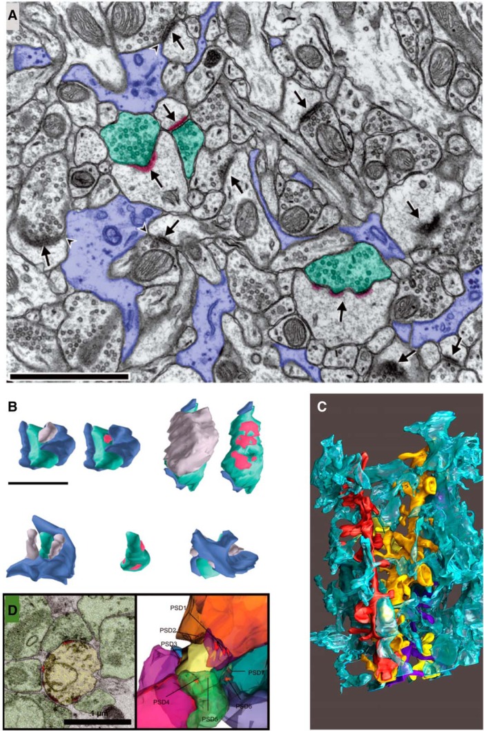
Figure 12.
Perisynaptic astroglial processes. A–C, Electron micrographs (A) and reconstructions (B, C) illustrate the nonuniform distribution of perisynaptic astroglial processes among synapses in mature hippocampal area CA1. Glial processes are lavender in A and B, and turquoise in C. Presynaptic axons are green, PSDs are red, and the postsynaptic spines are gray in A and B. Three dendrites (red, yellow, purple) are illustrated with the interdigitation of the glial processes in C. D, A multisynaptic dendritic spine, one of the few spines that remained in a surgically removed hippocampus from a patient with epilepsy. A and B are adapted from Ventura and Harris (1999); C is from Witcher et al. (2010).

