Figure 3.
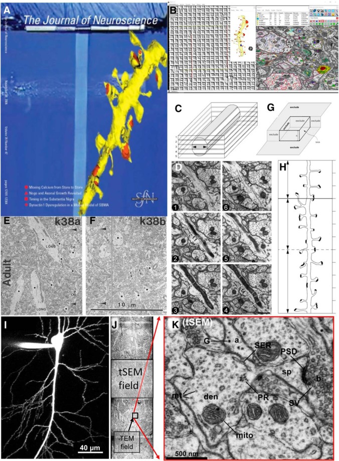
Interpreting quantitative outcomes from serial thin sections. A, JN cover that accompanied a Toolbox article covering material that was presented at two “Meet the Expert” sessions at the Society for Neuroscience meetings in 2005 and 2006 (adapted from Harris et al., 2006). B, Screenshot from Reconstruct, a tool for obtaining calibrated dimensions of objects reconstructed in 3D from serial thin sections (freely available for download with detailed manual at https://synapseweb.clm.utexas.edu/software-0). This image was generated from ongoing work in the Harris Laboratory. The windows show a calibration grid for determining the pixel size (left), a dendrite reconstructed using the Boissonnat surface tool along with a 1 μm scale cube made using the Box too (middle), and an EM image with defined objects (right). The objects list (top middle) outputs a .csv file with calibrated dimensions that can be imported into databases for statistical analyses, and there are drawing tools and color palettes (top right) to name and illustrate traces, stamps, and other defining objects (top right) that can be superimposed on aligned images. C, D, The cylindrical diameters method to estimate section thickness for accurate volume and surface area calculations. Adapted from Fiala and Harris (2001a). E, F, Neighboring fields from the same single section (see large arrowheads pointing to the same object on the left side of the line in F). Adjusting synapse density in neuropil areas or volumes requires subtracting areas or volumes of objects (black dots) that nonuniformly occupy space where synapses cannot form. Adapted from Harris et al. (1992). G, H, The unbiased brick (G) and unbiased dendritic length (H) methods for 3D stereology (from Fiala and Harris, 2001b). I, J, A CA1 pyramidal cell filled with Alexa Fluor dye at scale (I) next to three tSEM fields montaged across the apical CA1 dendritic arbor (J) to illustrate the large tSEM field size relative to the typical TEM field size. K, The resolution of the tSEM fields (≤2 nm pixel size) retains the same tissue elements as the original TEM fields. (I–K are adapted from Kuwajima et al., 2013a).

