Figure 6.
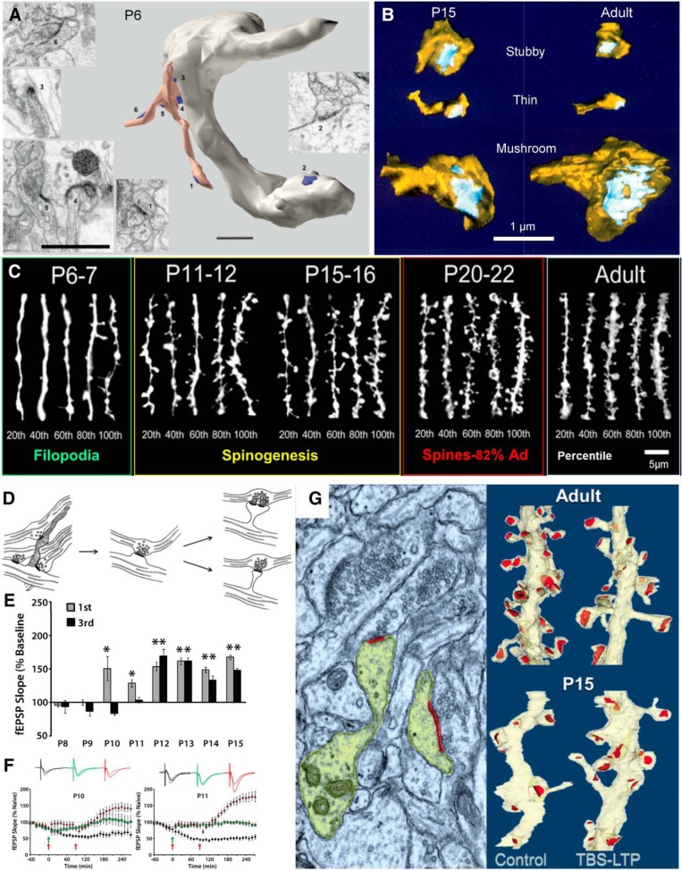
Synaptogenesis and the onset of LTP. A, EM sections and reconstruction of a varicose dendritic segment from CA1 stratum radiatum at P6. Multiple synapses (blue) are seen: one (2) is on the dendritic shaft and several are present near the tips and surrounding the base of a branched filopodium (peach colored). Adapted from Fiala et al. (1998). B, Dendritic spines of multiple shapes occur at both P15 and in young adults (P60–P70). From Harris et al. (1992). C, Confocal images of oblique dendrites from CA1 pyramidal cells in stratum radiatum, ranked by spine (protrusion) density across developmental ages compared with young adults (percentiles from 20 to 100 beneath each set of five dendrites. Adapted from Kirov et al. (2004a). By P20–P22, this density reaches ∼82% of the adult level (Ad). D, Model of spine outgrowth during development. Adapted from Harris (1999). E, Developmental onset of short-term potentiation (first hour) and late-phase LTP (third hour). F, At P10 and P11, a second bout of TBS (second red arrow) will induce late-phase LTP if sufficient time passes between the bouts, compared with slices that receive just one bout by TBS (green arrows and waveforms) or slices that undergo test pulse-induced depression (black waveforms and symbols). E and F are from Cao and Harris (2012). G, At P15, TBS induction of LTP produces new dendritic spines, the left side is colorized electron micrograph (yellow, dendrite and spine heads; red, postsynaptic density), and the right side is 3D dendritic segments from young adults (top) compared with P15 (bottom) that received control or TBS stimulation. From Watson et al. (2016). (A): Numbers 1, 3–6 match EMS of synpses on the branched filopodium. *p < 0.05, **p < 0.1.

