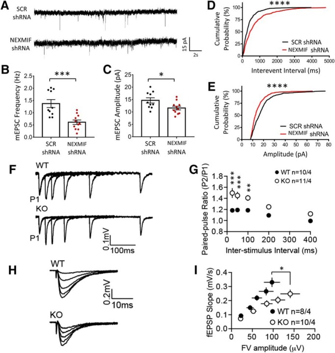
Figure 10.
Loss of NEXMIF results in altered synaptic transmission. A, mEPSC recordings from rat hippocampal neurons after infection with lentiviral scrambled (SCR shRNA) or NEXMIF shRNA, together with GFP. B, C, Quantification of mEPSC recordings showing a significant decrease in frequency and amplitude. D, E, Cumulative probability plots of mEPSCs showing a leftward shift in amplitude and a rightward shift in interevent interval for NEXMIF shRNA treated neurons. F, Representative traces from recordings performed in acute hippocampal slices from NEXMIF KO and WT mice to measure the paired-pulse ratio using an interstimulus interval of 25, 50, 100, 200, and 400 ms. G, Quantification of the paired-pulse ratio from recordings shown in I showed a significant increase in KO animals compared with WT controls (n = 10–11). H, Input/output curves from acute hippocampal slices from KO and WT hippocampal brain slices. Five stimulus intensities were chosen to elicit field excitatory postsynaptic responses. I, Quantification of the input/output curves showed that KO mice had a reduction in basal synaptic transmission compared with WT controls (n = 8–10). Data are represented as average ± SEM. *p < 0.05; **p < 0.01; ***p < 0.001; ****p < 0.0001.

