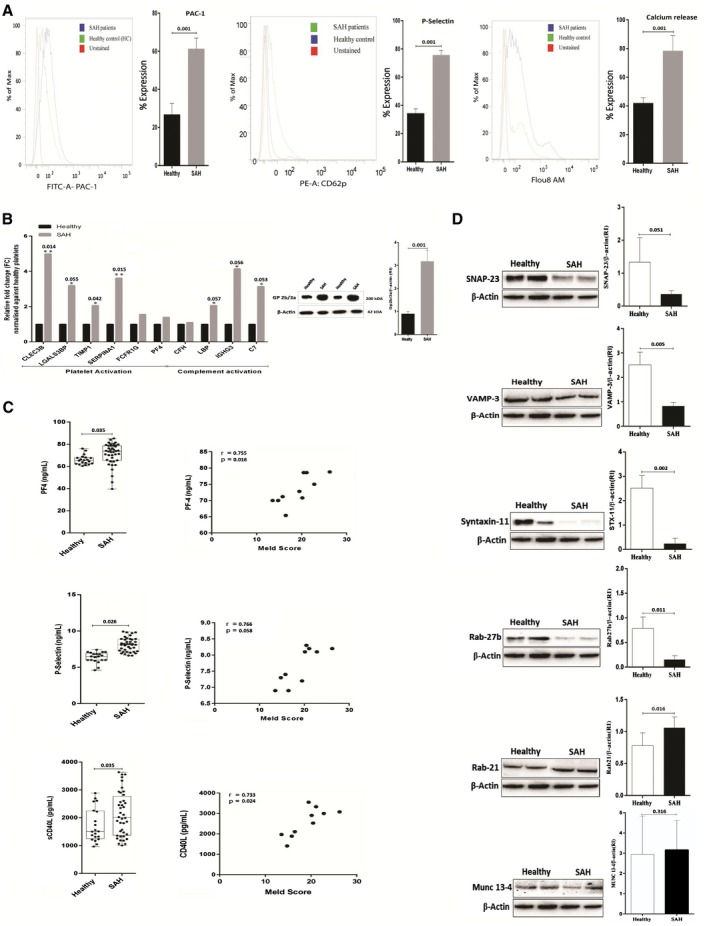
Figure 2.

Validation of platelet functionality in SAH. (A) Platelet activation/aggregation in SAH histogram shows the percent expression of PAC‐1, P‐selectin (CD62p), and intracellular Ca+2(secretion) in SAH and HC. (B) Increased expression (relative change) of various mRNA in SAH platelets linked to platelet and complement activation. Protein expression of Gp2b/3a/β‐actin ratio in platelets of patients with SAH. Results expressed as mean ± SEM (*P < 0.05, **P < 0.01 for SAH versus HC). (C) Patients with SAH showed higher plasma levels of PF‐4 (65.85 ± 0.9457 versus 81.13 ± 6.595 ng/mL, P < 0.02), P‐selectin (6.360 ± 0.1639 versus 10.118 ± 2.1492 ng/mL, P < 0.0001), and soluble cluster of differentiation 40 ligand (1,726 ± 134.4 versus 2,528 ± 1.525.4 pg/mL, P < 0.05). Circulating levels of PF‐4, P‐selectin, and sCD40L correlated with the MELD score in SAH (r > 0.5, P < 0.05). (D) Representative western blot of SNARE complex proteins in platelets isolated from SAH versus HC (n = 5). Results are expressed as mean ± SEM (*P < 0.05, **P < 0.01, ***P < 0.001 as indicated).
