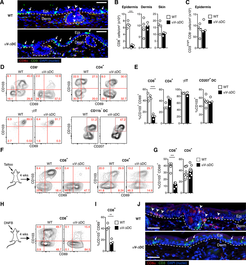
Fig. 1. Defective epidermal TRM cell formation in absence of αV integrins on DCs.
(A) Histological cross-sections from ear skin of 8–10 week-old αV-ΔDC and WT littermate control mice. Arrowheads indicate CD8+ CD3dim TRM cells in the epidermis of WT, but not αV-ΔDC mice, whereas arrows indicate CD3bright DETCs present in both strains. Dashed lines indicate the dermal–epidermal border. Scale bar = 50 μm. Epi: epithelium, Derm: dermis. (B, C) Density of CD8+ T cells in epidermis, dermis, and total skin (B) and of CD3bright CD8− T cells in epidermis (C). Data are means and replicates and representative of two independent experiments. Each symbol represents the mean of 3–4 values per animal obtained from different histological sections. (D, E) Expression of the tissue residence marker CD103 (as well as CD69 on T cells) on indicated immune cell subsets (Thy1+ CD8β+ T cells, Thy1+ CD4+ T cells, Thy1+ TCRd+ γΔT cells, CD11c+ MHC II+ CD11b− DCs) from skin of αV-ΔDC and WT littermate control mice. Data are means and replicates and representative of two independent experiments. (F-I) Expression of CD69 and CD103 on skin CD8+ or CD4+ T cells 4 weeks after sterile inflammation induced by mechanical irritation with a tattooing device (F, G) or topical DNFB treatment (H, I) in 8–10-week-old mice. Data are means and replicates and representative of five (F, G) or three (H, I) independent experiments. (J) Ear skin 4 weeks after DNFB treatment. Note the enrichment of CD8β+ CD3dim eTRM cells (arrow-heads) in WT mice, but their absence in the epithelium of αV-ΔDC, which is instead still populated by CD3bright DETC (arrows). Data are representative of two animals per group. Scale bar = 50 μm. **/***/****: p<0.01/p<0.001/p<0.0001 (Two-tailed unpaired Student’s t-tests in (B), (C), (E), (G), (I)).

