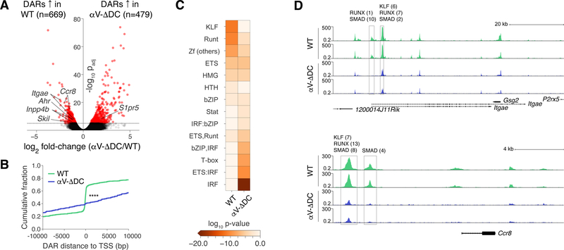
Fig. 4. Resting naive CD8+ T cells are epigenetically conditioned by TGF-β signals in secondary lymphoid tissues.
(A) Volcano plot of chromatin accessibility changes between CD44lo naive CD8+ T cells from WT and αV-ΔDC mice, considering merged DARs. (B) Cumulative distributions of distances from DARs in cells from WT (green) and αV-ΔDC (blue) animals to the closest transcription start site. The two-sample Kolmogorov–Smirnov was used to compare cumulative distributions. (C) Enrichment of indicated transcription factor binding motif families in DARs. (D) Normalized chromatin accessibility near the Itgae (top) and Ccr8 (bottom) loci. The rectangles mark detected DARs. All analyses were performed on 2 mice per group with similar results. ****: p<0.0001 (Two-sample Kolmogorov–Smirnov test in (B)).

