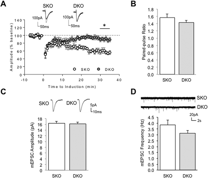
Figure 4.
Loss of mGluR-LTD at Parallel Fiber-PC synapses but Normal mEPSCs in Cerebellum of Grip1/2 DKO Mice. (A) Cerebellar LTD was induced by stimulating parallel fibers in conjunction with a PC depolarization at t =0 min. Evoked excitatory postsynaptic currents (EPSCs) were recorded from PCs in acute slices derived from one-month old Grip2 single KO (SKO) or neuron-specific Grip1/2 KO (Grip-DKO) animals. Depression was apparently observed in SKO (open circle) but not DKO (solid circle in grey). Example traces at 5-min before (thick line) and 25 min after (thin line) the LTD induction are overlaid on the top; Student’s t test; n=6, neurons from 4–5 mice per group. (B) Parallel fibers were stimulated with a paired pulse with a 100-ms interval. Paired-pulse ratio was calculated as EPSC2/EPSC1. There is no difference noted between SKO and DKO; Student’s t test; n=6 neurons from 4–5 mice per group. (C & D) Spontaneous AMPA receptor mediated miniature (mEPSC) were recorded in the presence of TTX and gabazine at −70 mV holding potential. AMPA mEPSC amplitude and frequency are comparable between PCs recorded from SKO and DKO mice. Individual mEPSCs and representative traces are showed on the top; Mann-Whitney test, n=17 and 20 from 5 pair of mice; *P<0.05.

