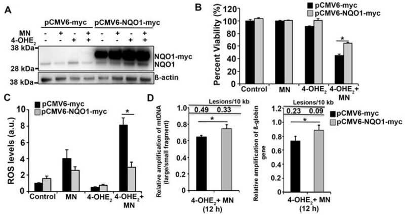
Fig. 4.
Overexpression of NQO1 reduces ROS formation and DNA damage induced by MN and 4-OHE2. A. Western blot analysis of plasmid expressed NQO1-myc and endogenous NQO1 levels in HCEnC-21T cells transfected with pCMV6-myc and pCMV6-NQO1-myc followed by treatment with 50 μM MN and 10 μM 4-OHE2. The bands corresponding to NQO1-myc and endogenous NQO1 are indicated. ß-actin serves as a normalizing control. B. Assessment of cell viability of HCEnC-21T cells transfected with pCMV6-myc (black) and pCMV6-NQO1-myc (grey) and treated with 10 μM MN and 20 μM 4-OHE2 for 24 h by Cell-titer glo assay. C. Determination of intracellular ROS production in HCEnCs transfected with vector (black) and NQO1 plasmid (grey) by DCFDA assay 5 h post treatment with 10 μM MN and 20 μM 4-OHE2. Statistical significance tested using Student’s t test. D. Assessment of mtDNA (left) and nDNA (right) damage by LA-qPCR analysis of HCEnC-21T cells transfected with vector (black) and NQO1 plasmid (grey) and treated with 10 μM MN and 20 μM 4-OHE2 for 12 h. (* p< 0.05, Student’s t test).

