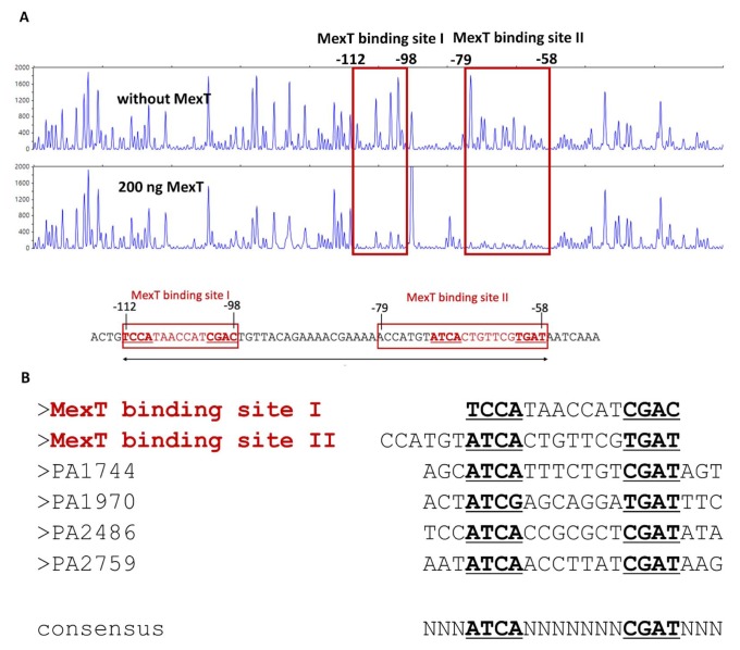
Fig. 5. MexT binding sequences identified by DNase I footprinting.
(A) A 630-bp DNA fragment containing the mexT-mexE intergenic region was labeled with 6-fluorescein amidite and then used as a DNA probe. The fluorescence-labeled DNA probe (400 ng) was incubated without or with 200 ng of purified FL MexT. The regions protected by MexT are indicated by red squares (MexT binding site I and II). Nucleotide numbers shown are the relative positions when the transcription start site mexE is designated as +1. (B) Comparison of the MexT-binding sequences. The two putative MexT-binding sequences in the mexEF-oprN regulatory region are shown at the top. The MexT-binding sequences from other promoters of MexT regulons are aligned below. Nucleotides in bold and underlined represent a partially conserved palindromic sequence, suggesting the potential consensus MexT-binding sequence, ATCA(N)7CGAT.

