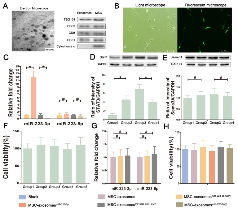
Fig. 1. Characterizations of exosomes derived from bone mesenchymal stem cells.
(A) The morphology of MSC-exosomes under an electron microscope and proteins expression in exosomes and MSCs. Arrows indicate exosomes as cup-shaped membrane vesicles of 40 to 100 nm in diameter. Western blot analysis indicates that exosomes are positive for CD9, CD63, CD81, and TSG101, but negative for cytochrome c. (B) The transfected cells under a light microscope and a fluorescent microscope at a magnification of ×200. Scale bars = 100 μm. After transfection, MSCs, which transfected successfully, displayed green fluorescence in fluorescent microscope image. (C) The expression levels of miR-223-3p and miR-223-5p in transfected MSC-exosomes. (D and E) The protein levels of Stat3 and Sema3A, the downstream target of miR-223-3p and miR-223-5p respectively, in transfected MSCs via Western blotting. Group 1, MSCsmiR-223-3p group; Group 2, MSCsmiR-223-3p-C°N group; Group 3, MSCsmiR-223-3p(i) group; Group 4, MSCsmiR-223-3p(i)-C°N group. (F) The effect of lentivirus, miR-223-3p knockin or knockdown on the proliferation viability of MSCs. Group 1, MSCs group; Group 2, MSCsmiR-223-3p-C°N group; Group 3, MSCsmiR-223-3p group; Group 4, MSCsmiR-223-3p(i)-C°N group; Group 5, MSCsmiR-223-3p(i) group. The results showed no difference. (G) The effect of lentivirus itself on the expression of miR-223-3p and miR-223-5p in MSC-exosomes. (H) The effect of MSC-exosomes and transfected MSC-exosomes on the viability of macrophages. The results showed no difference. Data are presented as mean ± SD from three independent experiments. *P < 0.05, #P > 0.05.

