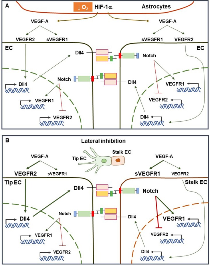
Fig. 1.
HIF-VEGF axis and Notch signaling cooperatively regulate endothelial specification. (A) In hypoxia regions, HIF-1α can be upregulated in ECs and astrocytes, leading to enhanced VEGF-A secretion. All ECs become activated by VEGF-A stimulation to express Notch and Dll4. (B) Notch signaling induces lateral inhibition and gives rise to nonuniform population of ECs in the presence of VEGF-A stimulation. Stalk cells are subject to enhanced Notch signaling, which represses VEGFR2 transcription, while stimulating the expression of the decoy receptor, soluble VEGFR1 (sVEGFR1). Tip cells receive a low Notch signal, allowing for high VEGFR2 and low sVEGFR1.

