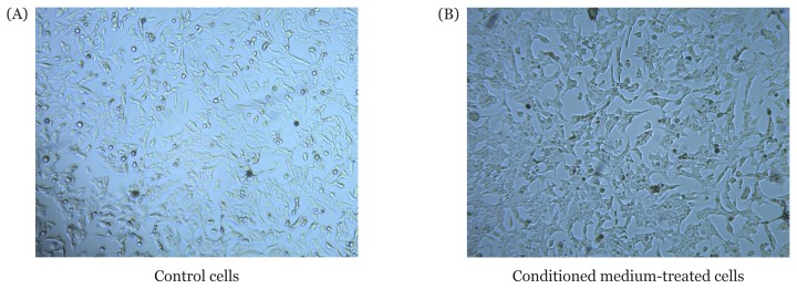
Figure 1.
T98G cell morphology. 4×10⁵ cells were seeded in triplicate on a 12-well plate in high-glucose Dulbecco’s Modified Eagle’s Medium with 10% FBS, 1% amphotericin and 1% streptomycin-penicillin at 37 °C in 95% O2 and 5% CO2. The following day, the medium of treated cells was replaced with 50% (v/v) conditioned medium of UCMSC-CM, whereas the medium of the control cells was replaced with 50% (v/v) minimum essential medium alpha. Both cultures were further incubated for 24 h. Cell morphology was then observed using an inverted microscope (100× magnification)

