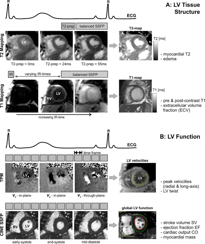
Figure 2:
Combined T2 mapping, T1 mapping, tissue phase mapping (TPM), and cine steady-state free precession (SSFP) MRI for the comprehensive evaluation of myocardial, A, tissue structure and, B, function. T2 mapping is based on diastolic balanced SSFP imaging with different T2 preparation times. T1 mapping uses balanced SSFP imaging with different inversion recovery (IR) times. TPM is based on electrocardiographically (ECG) gated black-blood prepared phase contrast MRI with three-directional velocity encoding. T2 mapping, T1 mapping, and TPM were acquired in short-axis orientation (base, mid, and apex) during breath holding. CO = cardiac output, EF = ejection fraction, LV = left ventricular, prep = preparation time, RV = right ventricular, SV = stroke volume.

