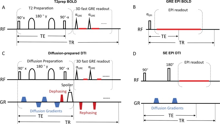
Figure 1:
Image shows pulse sequence diagrams of, A, three-dimensional (3D) T2-prepared (T2prep) blood oxygenation level–dependent (BOLD) functional MRI, B, conventional two-dimensional multislice gradient-echo (GR) echo-planar imaging (EPI) BOLD functional MRI, C, 3D diffusion-prepared diffusion tensor imaging (DTI), and, D, conventional 2D multislice spin-echo (SE) EPI DTI. One entire image volume was acquired in a single repetition time (TR) period in all sequences to avoid well-known phase errors in multishot approaches. Details of these pulse sequences are described in Appendix E1 (online). RF = radiofrequency, TE = echo time.

