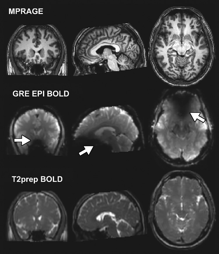
Figure 3:
Representative anatomic (magnetization-prepared rapid acquisition gradient-echo [MPRAGE]), gradient-echo (GRE) echo-planar imaging (EPI) blood oxygenation level–dependent (BOLD), and T2-prepared (T2prep) BOLD images acquired in participant wearing titanium dental brace at 3.0 T. Image plane from left to right: coronal, sagittal, and axial. Signal dropouts can be seen on EPI-based images in regions close to brace (arrows). T2-prepared BOLD images were not affected by strong susceptibility effect.

