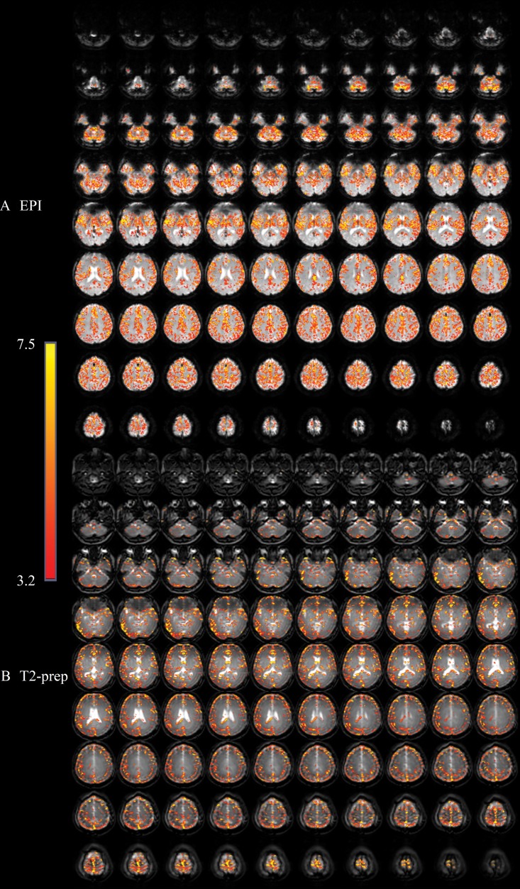
Figure 4:
Activation maps overlaid on original functional MRI scans from, A, gradient-echo echo-planar imaging (EPI) and, B, T2-prepared (T2prep) blood oxygenation level–dependent functional MRI approaches, respectively, in axial imaging plane from healthy participant wearing titanium dental braces during breath-hold task. Activated voxels are highlighted with their corresponding t scores from general linear model analysis with identical statistical threshold. Range of t scores is indicated by scale bar.

