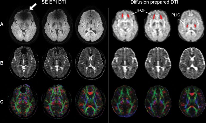
Figure 5:
Spin-echo (SE) echo-planar imaging (EPI) and diffusion-prepared diffusion tensor imaging (DTI) axial images acquired at 3.0 T in participant wearing metallic dental braces. A, Raw diffusion-weighted images, B, calculated apparent diffusion coefficient maps, and, C, fractional anisotropy map color coded by V1 (principal eigenvector) orientation (standard red, green, and blue convention). Susceptibility artifacts were observed on SE echo-planar image in regions close to braces (arrow). No obvious artifacts were seen on diffusion-prepared diffusion tensor image. Regions of interest of inferior fronto-occipital fasciculus (IFOF) and posterior limb of internal capsule (PLIC) used in subsequent quantitative analysis are highlighted on diffusion-prepared diffusion tensor images with red.

