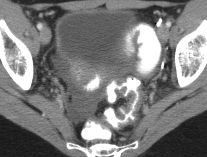
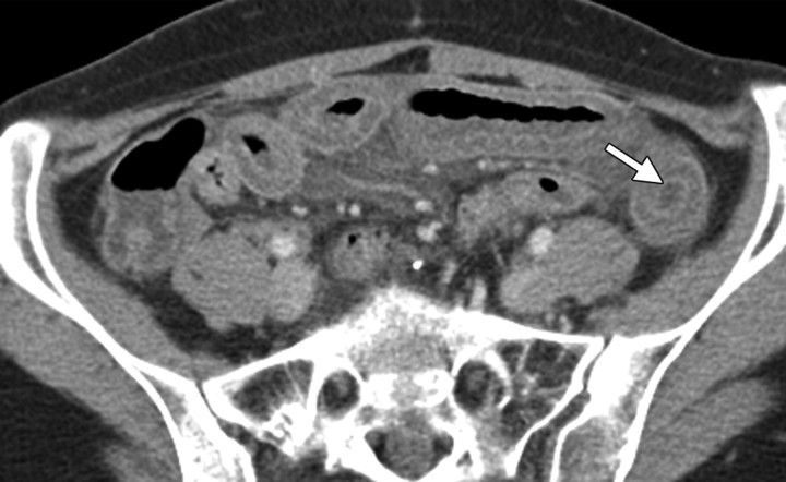
Enteritis in a 27-year-old woman who had undergone total abdominal hysterectomy, chemotherapy, and radiation therapy for metastatic small cell adenocarcinoma of the cervix. (a) Follow-up CT scan obtained after the administration of intravenous and oral contrast material shows a thick-walled sigmoid colon and rectum, as well as free fluid in the pelvis. (b) CT scan from the same examination shows an abnormal thick-walled small bowel with enhancing mucosa (arrow), abnormal findings that are consistent with enteritis but do not indicate a malignant small bowel process.

An official website of the United States government
Here's how you know
Official websites use .gov
A
.gov website belongs to an official
government organization in the United States.
Secure .gov websites use HTTPS
A lock (
) or https:// means you've safely
connected to the .gov website. Share sensitive
information only on official, secure websites.

