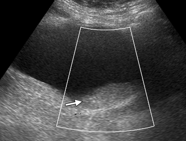
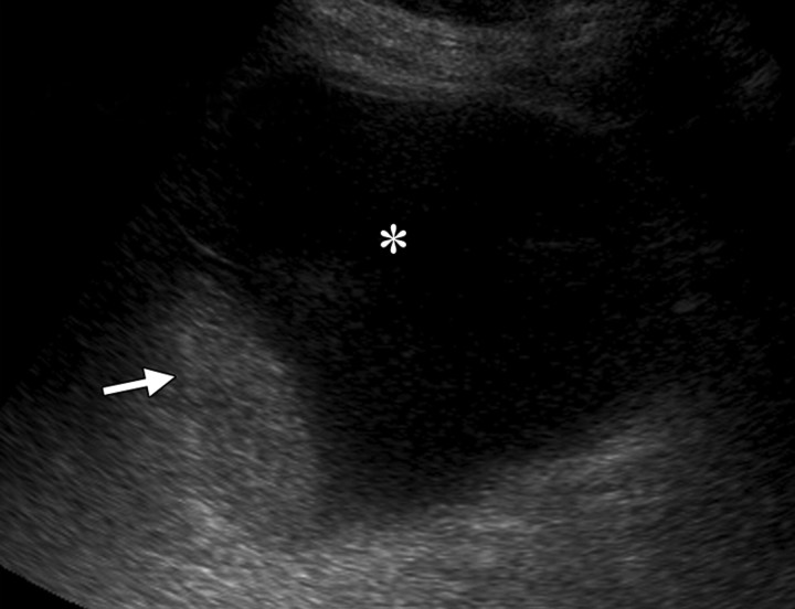
Mobile clot in a 51-year-old woman who had undergone radiation therapy for cervical carcinoma. The patient presented with frank hematuria. (a) Transabdominal Doppler US image (shown in black and white), obtained with the patient supine, demonstrates an echogenic filling defect (arrow) in the urinary bladder. No flow was evident. (b) US image obtained with the patient lying on her right side shows an echogenic mass (arrow) in a dependent position within the urinary bladder (*), a finding that is consistent with a mobile clot.

An official website of the United States government
Here's how you know
Official websites use .gov
A
.gov website belongs to an official
government organization in the United States.
Secure .gov websites use HTTPS
A lock (
) or https:// means you've safely
connected to the .gov website. Share sensitive
information only on official, secure websites.

