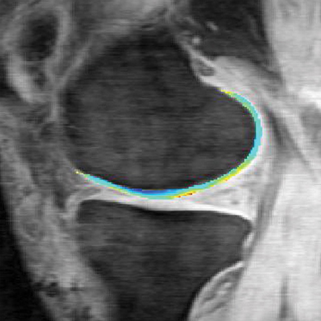
T2 maps obtained in knee cartilage in a 51-year-old man after anterior cruciate ligament repair show higher T2 values (yellow) in the medial (a) and lateral (b) tibiofemoral compartments, findings indicative of deterioration of the integrity and orientation of the collagen network. Maps were obtained at 3.0 T with a sagittal 3D T2-weighted multiecho SE sequence (TR/TE = 9.3/3.1, 13.5, 23.9, 44.8; recovery time = 1500 msec; field of view = 14 cm; matrix = 256 × 192; section thickness = 3 mm; bandwidth = 31.25-kHz). Scale is in milliseconds.

An official website of the United States government
Here's how you know
Official websites use .gov
A
.gov website belongs to an official
government organization in the United States.
Secure .gov websites use HTTPS
A lock (
) or https:// means you've safely
connected to the .gov website. Share sensitive
information only on official, secure websites.

