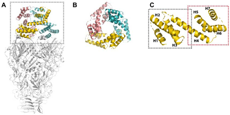
Figure 3. Structure of the N-Helical-lid.
(A) Side view of a PdpA trimer, with its N-helical lid colored and enclosed within the dotted box.
(B) Top view of the trimeric N-helical lid.
(C) Ribbon diagram of a monomeric unit of the trimeric N-helical-lid. The regions of residues 1-75 and 70-169 are marked by a grey and red dashed box, respectively. The 7 helices present in the N-helical-lid are labeled as H1 to H7.

