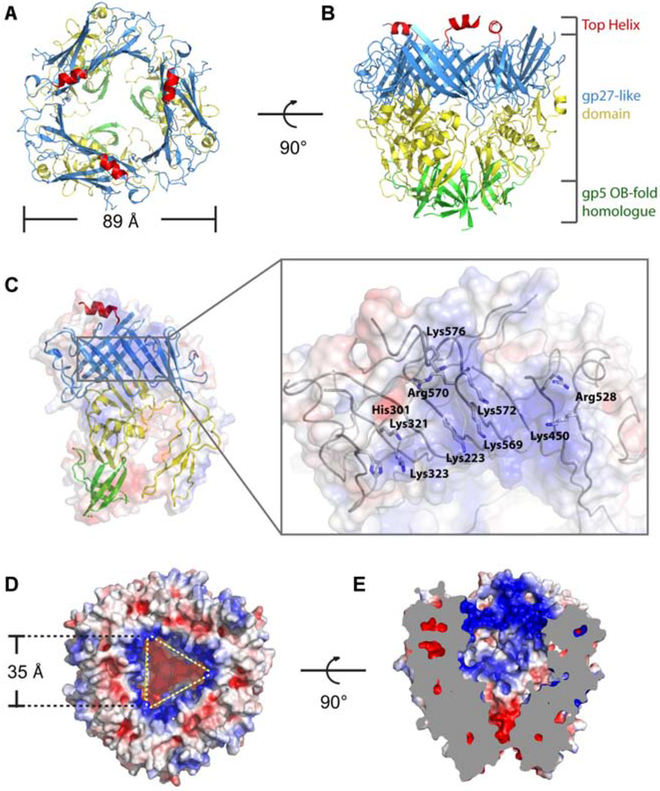
Figure 4. Structure of the head.
(A, B) Top (A) and side (B) views of the head. The top helix, upper ring, middle ring and lower ring are colored red, blue, yellow and green, respectively. (C) Electrostatic surface representation of the monomeric head domain showing the inner surface of the funnel. The electrostatic surface is shown semi-transparently, superposed with the ribbon diagram of the atomic model. Inset: atomic model of the positive charged “belt” region is shown as grey wires with key residues labeled. (D, E) Top (D) and side cut-away (E) views of electrostatic surface potential distribution of the head showing the cavity. See also Video S3.

