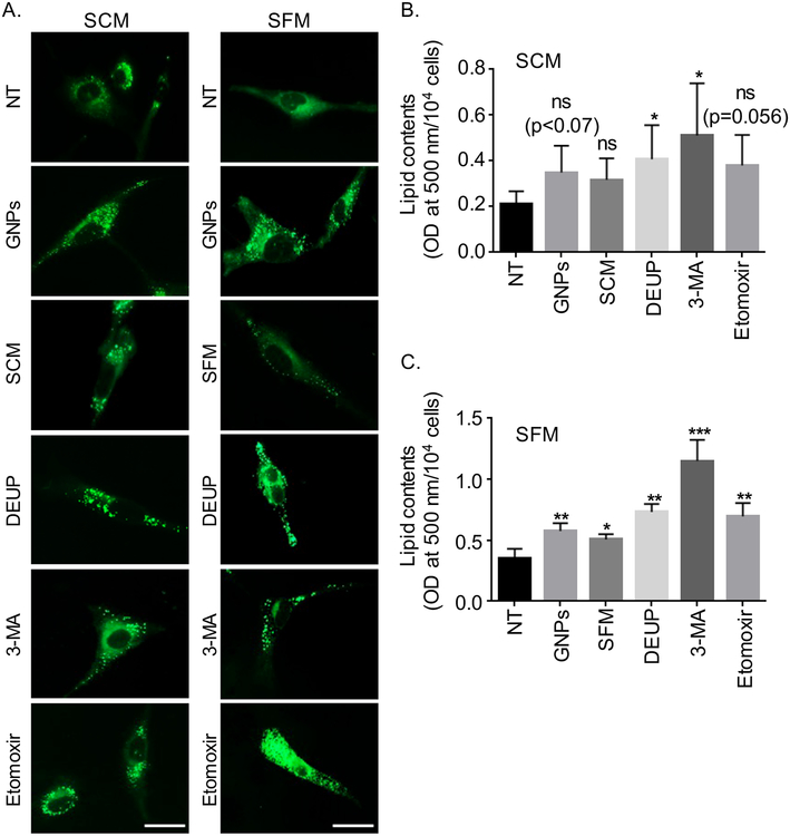
Figure 4.
Utilization of lipid droplets by CAFs. (A) CAFs were treated with GNPs or remained untreated (NT) for 48h. GNPs-treated CAFs were further incubated with SFM, SCM, DEUP (50μM), 3-MA (5 mM), etoximir (50 μM) either in SCM or SFM for 24h or remained untreated after which LDs were visualized by BODIPY fluorescence (green, λex./em.=493/503 nm). LDs were stained with BODIPY for 30 mins, fixed and then visualized under fluorescence microscope. Representative images of cells were shown here from three independent experiments (n=3). Scale bar 20 μm. (B-C). Quantification of lipid contents in CAFs using Oil Red O staining. Experiments were performed as above except staining with Oil O Red instead of BODIPY. The optical density (OD) value of Oil Red O dye was measured at 500 nm and was expressed as OD/104 cells. Statistical significance was determined by student’s unpaired t-test of three independent experiments (mean±s.d., n=3, ns=non-significant, *p<0.05, **p<0.01 and ***p<0.001, NT vs treated groups).

