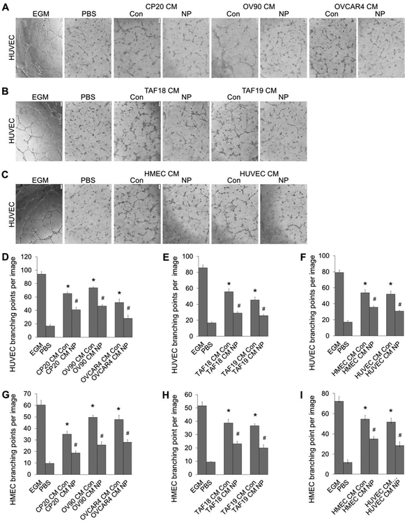
Figure 3.
Tube formation of EC treated with CM from CCs, CAFs or ECs. CM were diluted with equal volume of fresh EBM before use. HUVECs or HMECs were starved in EBM for 16 h before trypsinized and incubated for 30 min with the CM. ECs were then seeded 20,000 cells/well (HUVEC) or 30,000 cells/well (HMEC) to 96-well plate coated with 50 μl Matrigel (1:1 diluted with EBM). Images of tubular network were taken 4 h later. Tube formation was evaluated by counting the branching points of the tubular network with ImageJ. EGM or PBS diluted with equal volume of EBM was used as positive or non-treatment control. (A-C) Typical images of tube formation of HUVECs treated with CM of CCs, CAFs or ECs. (D-F) Quantification of HUVEC tube formation. (G-l) Quantification of HMEC tube formation. Experiments were performed in triplicate and repeated 3 times with similar results. Con: control, NP: AuNPs. Scale bar: 100 μm. *, p<0.05, compare to PBS: #, p<0.05, compare to corresponding Con.

