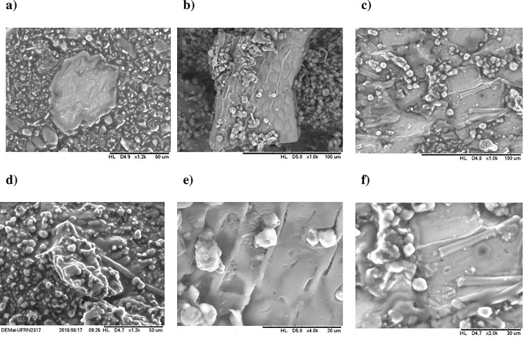
Fig 1.
Scanning electron microscopy of Cantaloupe melon (Cucumis melo L. var. reticulatus) seed flour: (a) 1,2 k, (b) 1,0 k, (c) 1,0 k, (d) 1,2 k, and 4,0 k, and (f) 2,0 k. Fig 1e represents two configurations; the heaviest particle indicates a higher atomic number; therefore, it has a stronger glow (clearest picture). In contrast, the lighter elements (lower atomic numbers), stand out because they are darker.

