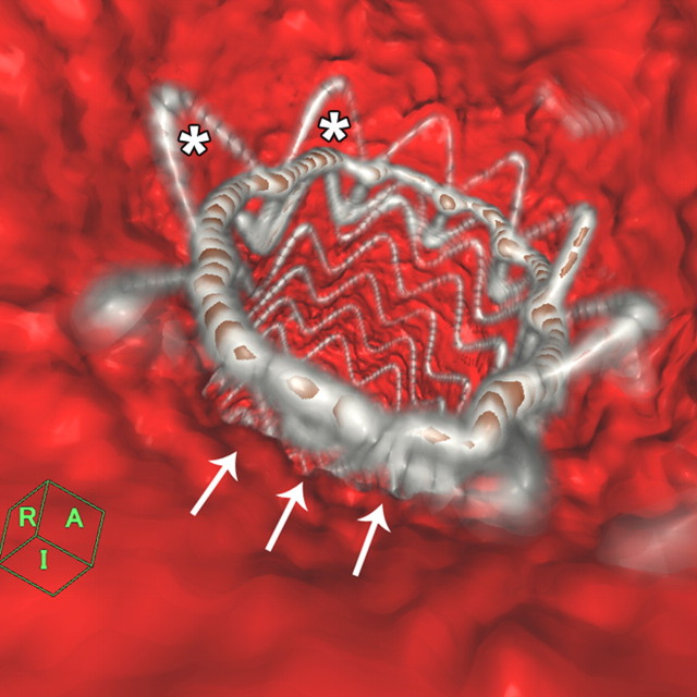
Figure 1b:
Images obtained in an 84-year-old woman who underwent TEVAR for an atherosclerotic aortic aneurysm show bird-beak configuration resulting in type Ia endoleak. (a) Thin-slab maximum intensity projection shows bird-beak configuration (arrowhead)—imperfect apposition at proximal end of stent-graft to lesser curve of aortic arch—resulting in wedge-shaped gap between undersurface of the stent-graft and aortic wall. Length (two-headed arrow) and angle of the bird-beak were measured with three-dimensional workstation functions. Scalloped flares (small arrows) at the proximal end of the device were excluded from measurement of bird-beak length. Leakage of contrast medium is observed flowing continuously from the bird-beak into the aneurysmal sac, signifying type Ia endoleak (large arrow). (b) Virtual angioscopic CT image shows proximal end of stent-graft, as viewed from ascending aorta. Proximal end of stent-graft is incompletely attached to aortic wall, resulting in a gap between the undersurface of the stent-graft and the aortic lesser curve wall (arrows). Scalloped flares along greater curvature of device are well apposed to aortic wall (*).

