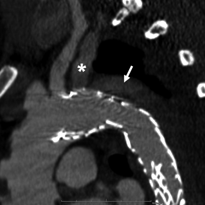
Figure 2a:
Images obtained in a 42-year-old man who underwent TEVAR for subacute type B aortic dissection show bird-beak configuration and developed a complex primarily type IIs endoleak. (a) Oblique sagittal thin-slab maximum intensity projection image shows enhancement of aortic false lumen (arrow), apparently fed by left subclavian artery (*). Subsequent catheter angiography revealed small type Ia endoleak with anterograde flow from the aortic arch into the LSCA and retrograde flow in the aortic false lumen into the LSCA, which had to-and-fro flow. The dominant mechanism of endoleak was type IIs, as contrast enhancement in the left subclavian artery was almost the same as that in adjacent areas of endoleak, gradually fading away in the distal part, and false lumen near the proximal end of the stent graft was less enhanced than that in aortic true lumen. (b) Thicker-slab maximum intensity projection image shows bird-beak configuration (arrowhead). Stent-graft showed severe infolding at mid- and distal portions because of constraint of the device within narrow and tapered true lumen.

