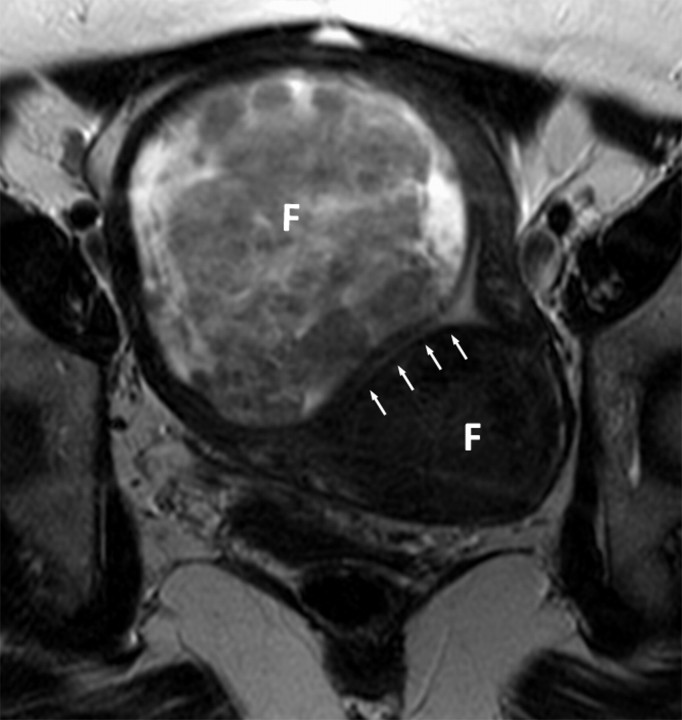
Figure 1a:
(a, b) Oblique coronal T2-weighted MR images (6317/106) of a 48-year-old woman with adenocarcinoma cells of indeterminate primary origin at cervix biopsy. A grossly enlarged uterus was noted clinically, raising suspicion of a corpus tumor with cervical extension. MR images show large fibroids (F) compressing the endometrial cavity. Endometrial stripe (arrows) was normal in thickness (4 mm) and signal intensity. A cervix tumor (arrowheads) was identified as an area of intermediate signal intensity extending into cervical stroma. Final pathologic diagnosis from the hysterectomy specimen was a stage T1b1 endocervical adenocarcinoma.

