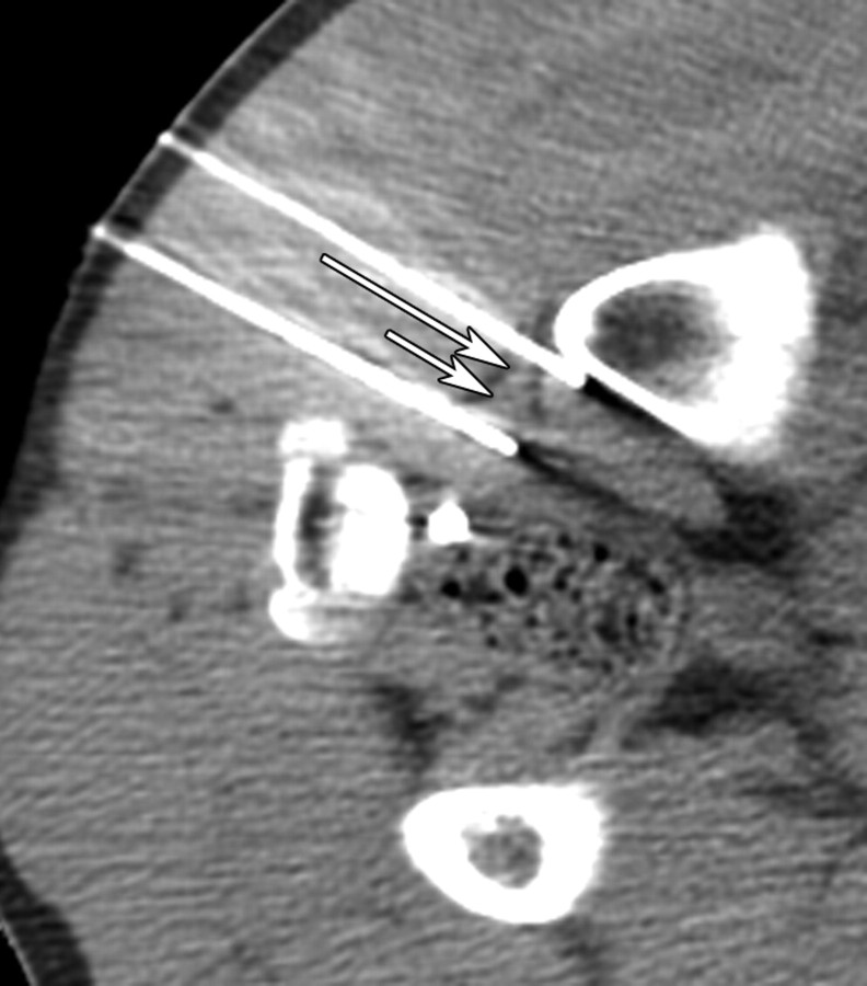
Figure 1b:
(a) CT image before IRE ablation shows level of planned probe entry with right sciatic nerve (long arrow) and accompanying arteria comitans nervi ischiadici (short arrow) surrounded by fat tissue. (b) CT image after placement of IRE electrodes with right sciatic nerve (long arrow) and accompanying artery (short arrow) between electrode tips.

