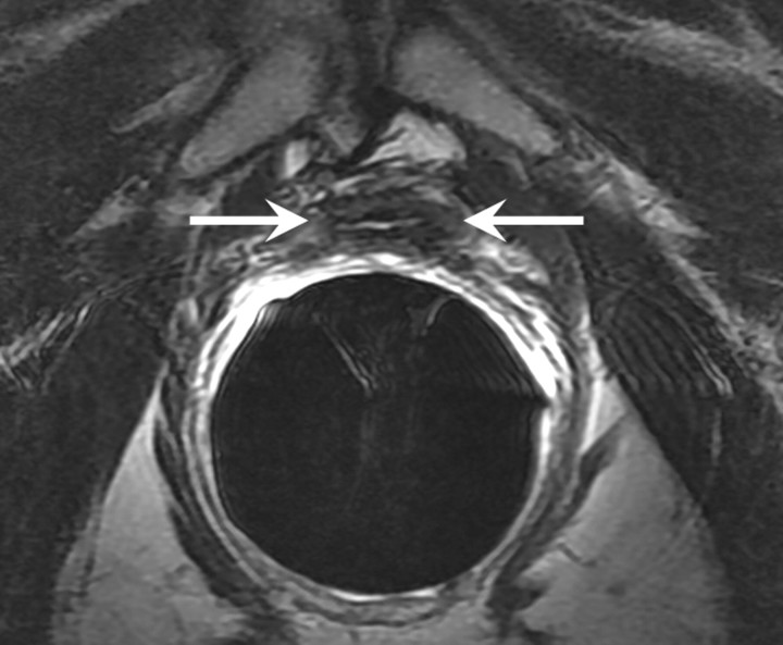
Figure 1a:
T2-weighted MR images in 74-year-old man who has undergone prostatectomy. (a) Transverse and (b) sagittal images show that urinary bladder neck is pulled down and anastomosed to the membranous urethra. The normal vesicourethral anastomosis (arrows) has a uniform shape, with low signal intensity, similar to that of the urinary bladder wall, on T2-weighted images.

