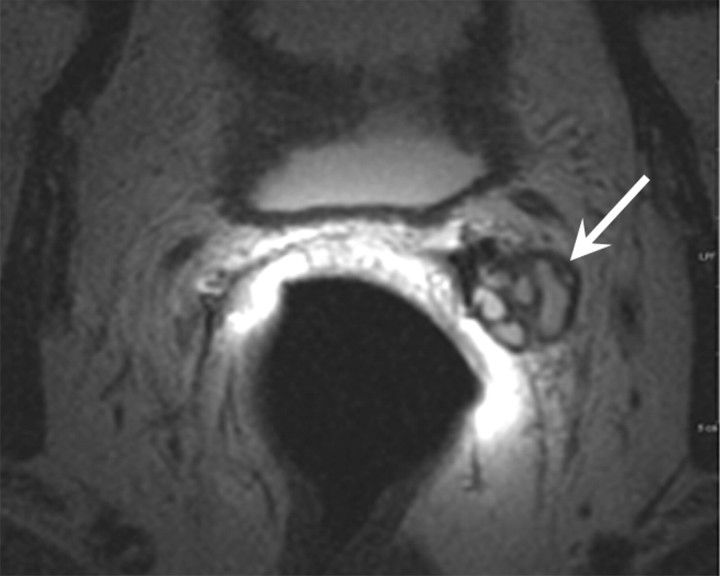
Figure 2b:
Retained seminal vesicle (arrow) is seen on transverse (a) CT and (b) MR images in 64-year-old man who has undergone prostatectomy. Note that it is difficult to characterize the “lesion” (arrow) as a retained seminal vesicle on a. However, b clearly shows the typical convoluted tubular appearance of the seminal vesicle.

