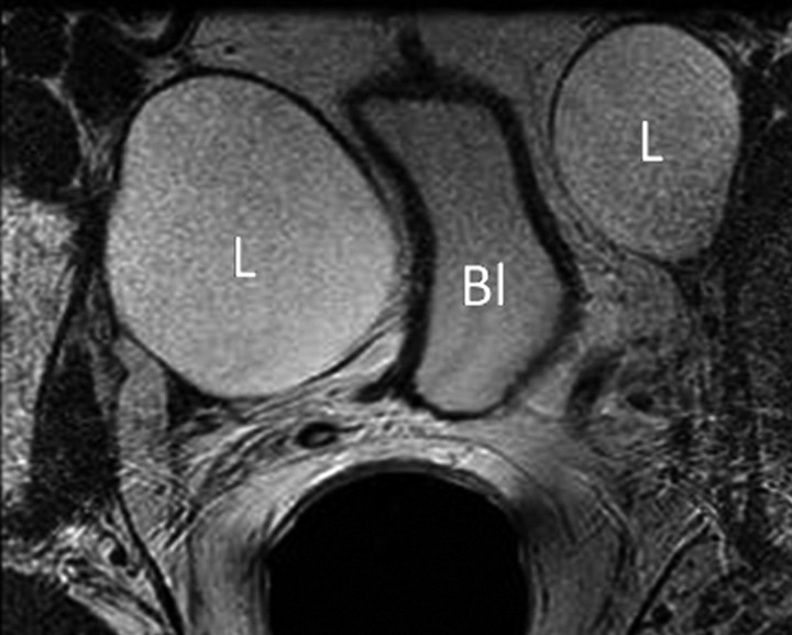
Figure 3a:
Transverse MR images in three patients who have undergone prostatectomy. (a) T2-weighted image shows bilateral pelvic sidewall lymphoceles (L) that compress the urinary bladder (Bl). (b) T2-weighted image shows defect in posterior urinary bladder wall (arrow) just above the level of the vesicourethral anastomosis with an adjacent urinoma (∗). (c) T2-weighted and (d) contrast-enhanced T1-weighted images show abscess (A) with thick irregular walls in prostatectomy bed.

