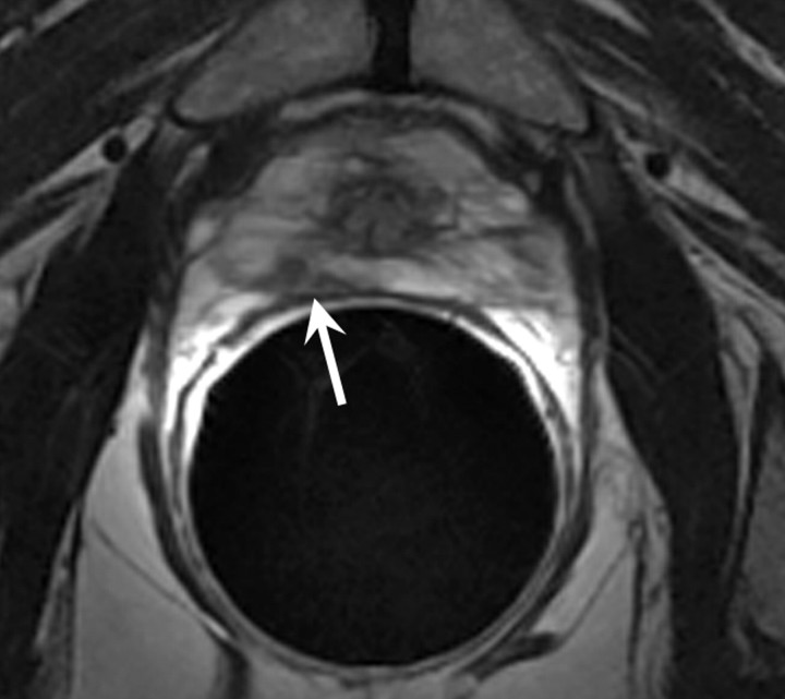
Figure 9a:
Transverse T2-weighted MR images obtained (a) before and (b) 2 years after external-beam RT. (a) Focal tumor (arrow) is shown in right peripheral zone at apex of the prostate. (b) Recurrent mass with extracapsular extension (arrow) can be seen at site of original tumor, which was biopsy-proved prostate cancer recurrence.

