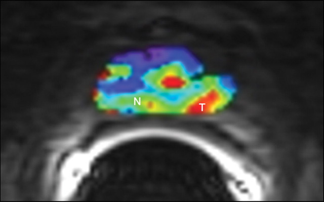
Figure 11b:
Biopsy-confirmed prostate cancer recurrence in left peripheral zone in 72-year-old patient after external-beam RT. (a) Transverse T2-weighted MR image shows area suspicious for tumor recurrence (T), but the possibility of normal posttreatment change cannot be excluded. (b) Ktrans map from axial dynamic contrast-enhanced MR imaging shows increased enhancement in area suspicious for tumor recurrence (T), compared with area of normal prostate tissue (N). (c, d) Contrast enhancement–time curves generated from dynamic contrast-enhanced MR imaging data show rapid avid enhancement in (c) region suspicious for tumor, as compared with (d) normal prostate tissue.

