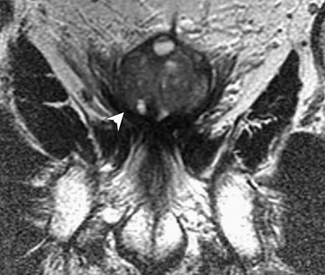
Figure 17b:
(a) Transverse and (b) coronal T2-weighted MR images in 59-year-old patient treated with high-intensity focused ultrasound shows thickened prostatic capsule (arrow) and extensive tissue fibrosis around the prostate (arrowhead). There is diffusely decreased volume in the peripheral zone with benign prostatic hyperplasia in the transition zone.

