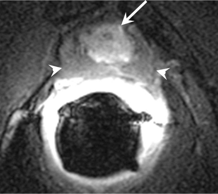
Figure 18b:
Transverse (a) T2-weighted and (b) contrast-enhanced T1-weighted MR images in 56-year-old patient with increasing PSA level after cryotherapy show recurrent tumor (arrow) Note that tumor has intermediate signal intensity on a and is markedly enhanced on b. Postcryotherapy fibrosis (arrowheads) demonstrates low-signal-intensity on a and does not enhance on b.

