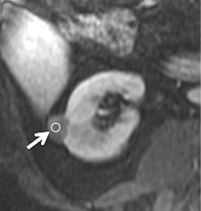
Figure 4c:
Axial gradient-echo fat-suppressed T1-weighted images (a) before and (b–d) after the administration of gadolinium-based contrast material in the (b) corticomedullary, (c) nephrographic, and (d) excretory phases of enhancement in a 69-year-old male patient demonstrate a mildly enhanced exophytic right renal mass arrow). The %SI changes from the precontrast to the corticomedullary (69%), nephrographic (118%), and excretory (108%) phases (averaged for all readers) were observed. The final histologic finding was papillary carcinoma.

