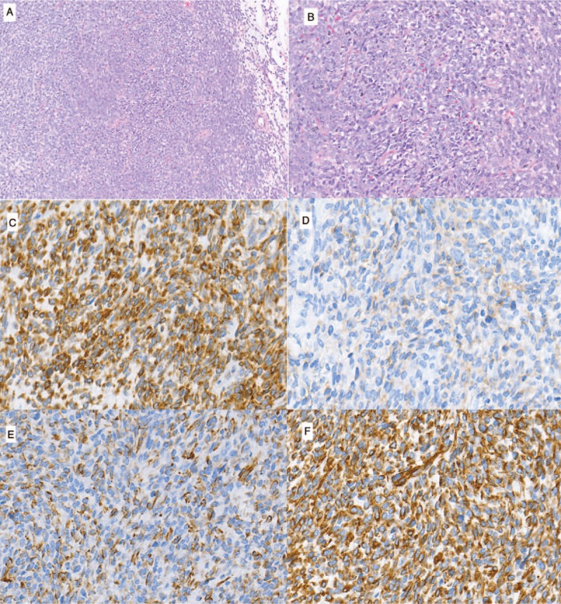
Figure 3.

An analysis of the surgical specimen revealed a spindle cell tumor (A and B). A: Hematoxylin and Eosin (H&E) staining (100 × ), A2: H&E staining (200 × ). The immunohistochemical staining was positive for Bcl-2 (C) and CD99 (D) and WT-1(E) and vimentin(F) (400X).
