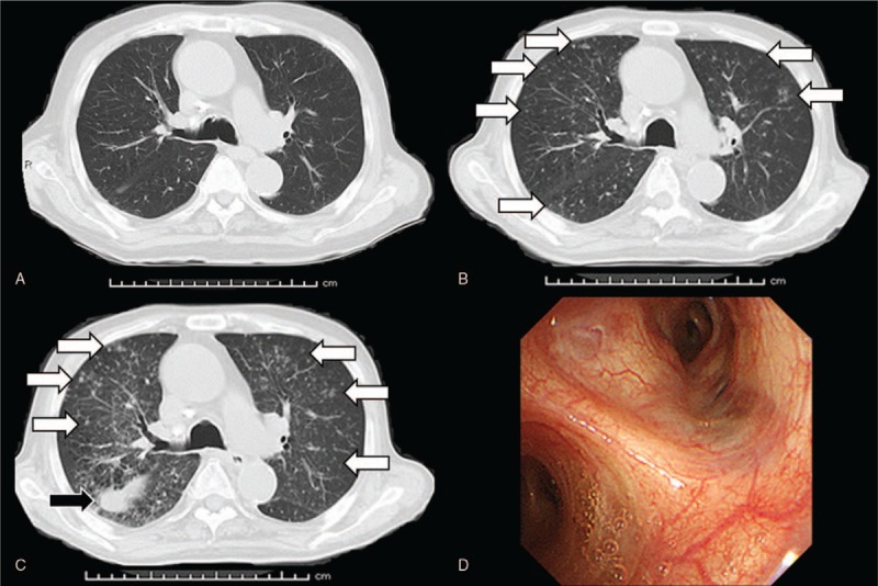
Figure 1.

Compared with the findings of chest computed tomography before cabazitaxel treatment (A), small nodules distributed around the bilateral lungs (white arrows) emerged 15 days after the third course of cabazitaxel therapy (B). Findings of diffusely distributed small nodules worsened (white arrows) and pleural effusion (black arrow) was newly observed 23 days after the third course of cabazitaxel therapy (C). Bronchoscopy of the respiratory system did not yield any significant findings (D).
