Figure 4.

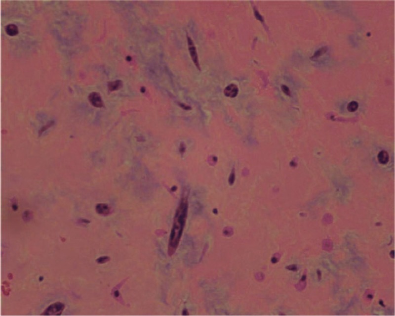
Histology of the excised tumor. The tumor consists of an acid-mucopolysaccharide-rich stroma. Polygonal cells with scant eosinophilic cytoplasm can be observed in the matrix. Hematoxylin–eosin stain.

Histology of the excised tumor. The tumor consists of an acid-mucopolysaccharide-rich stroma. Polygonal cells with scant eosinophilic cytoplasm can be observed in the matrix. Hematoxylin–eosin stain.